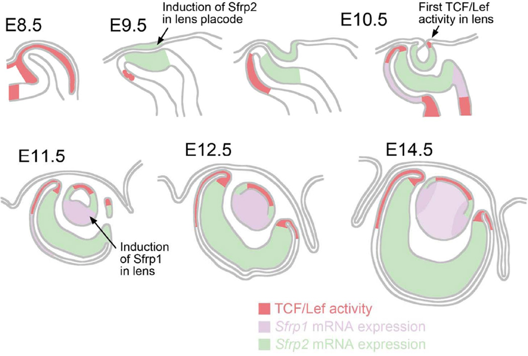
Fig. 8. Non-overlapping distribution of Sfrp1 and Sfrp2 expression and TCF/Lef activity.
Sfrp2 mRNA is detected in developing lens cells at lens placode (E9.5) and lens pit stages (E10.5). It becomes restricted to a band of cells above the lens equator at E11.5 and is not detectable in lens after E16.5 (see Chen et al, 2004). Sfrp1 expression is not detected in lens cells until the lens pit stages and then becomes strongly expressed in the elongating primary fibers of the lens vesicle at E11.5. Strong expression persists in fibers through all embryonic stages with slightly more prominent expression detected in cortical fibers. TCF/Lef activity is first detected at the ventral edge of lens pit at E10.5 and then found in the central lens epithelial cells after E11.5. The number of cells showing TCF/Lef activity in the lens epithelium diminishes after E16.5. Sfrp2 expression and TCF/Lef activity show complementary patterns in the developing retina. Although Sfrp1 and Sfrp2 do not show overlapping patterns of expression, it is only in DKOs of Sfrp1 and Sfrp2 that abnormal development of both lens and retina is evident. Moreover, analysis of DKOs reveals that Sfrp1 and Sfrp2 do not have a suppressive effect on TCF/Lef activity in eye but rather appear to act as enhancers of TCF/Lef activity in adjacent cells.

