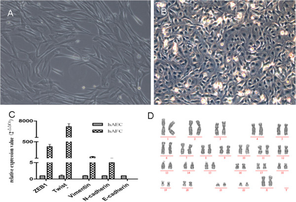
Figure 4.

Characterization of cell type in a human amniotic fluid cell (hAFC) line grown from one sample obtained via amniocentesis. (A) The hAFCs displayed morphology typical of mesenchymal cells. (B) Human amnion epithelial cells (hAEC) grown in culture for comparison. (C) Comparison of relative gene expression levels for mesenchymal and epithelial markers relative to 18S RNA; Compared with hAECs, hAFCs cells expressed high levels of mesenchymal markers, such as zinc finger E-box binding homeobox 1 (ZEB1), Twist, Vimentin and N-cadherin; conversely, hAFC did not express the epithelial marker E- cadherin. (D) Karyotype analysis showed a normal chromosomal complement (46,XX) in hAFCs grown herein. Magnification, 100x (A), 200x (B).
