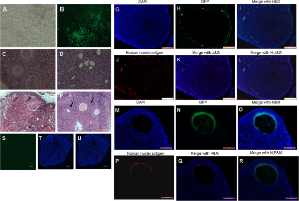
Figure 5.
Transplantation of a line of GFP-transfected human amniotic fluid-derived cells (hAFCs) into chemotherapy-sterilized recipient mice. (A) hAFC cell line derived from clone cells grown to 85% density. (B) GFP-transfected hAFCs. (C-F) Representative H&E micrographs of ovary sections from: (C) non-sterilized normal control mice. (D) Sterilized non-transplanted mice after a 2 month-recovery period showing stroma, and atretic primordial or primary follicles. (E, F) sterilized recipient mice following transplantation of hAFCs; arrows indicate follicles at various stages of maturational development. Sections display primordial (E), as well as primary and large antral follicles (F). (G-R) Immunofluorescence of ovarian sections from sterilized mice 2 months after transplantation with GFP-transfected hAFCs. (G-I) GFP staining is observed in ovarian stroma. (M-O) Follicles containing GFP-positive (green) oocytes. Blue: DAPI immunofluorescence. (S-U) Culture-medium injected ovaries in control sterilized mice lack GFP signal following a 2-month recovery period. (G-R) Double-staining with GFP and human nuclei antigen were performed to look for the derivation of GFP positive cells in recipient ovaries 2 months after hAFCs transplantation. (G-L) Ovarian sections stained with GFP and anti–human nuclei antibody revealing grafted hAFCs in stroma. (M-R) GFP staining was co-locolized with human nuclei antigen in antral follicles of recipient ovaries after hAFCs transplantation for 2 months. Scale bars = 50 μm (A, B, D, E, S-U); 100 μm (G-R); 200 μm (C, F).

