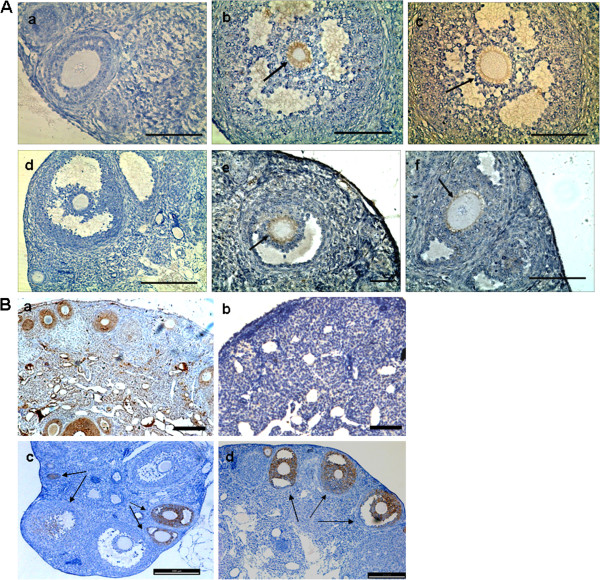
Figure 6.

Grafted hAFCs infiltrate the chemically-damaged murine ovarian tissue and restore ovarian function. (A) Immunohistochemistry for human-specific antigens detecting grafted cells resulting from transplantation of human amniotic fluid cells into the ovaries of chemically-sterilized mice. (a) Sample for human nuclei antigen-negative ovarian section following hAFC transplantation. (b, c) Human nuclei antigen expression in recipient ovaries 2 months after hAFCs transplantation. (d) Sample for human follicle stimulating hormone receptor (FSHR)-negative ovarian section following hAFCs transplantation. (K, L) Human FSHR expression in recipient ovaries 2 months after hAFCs transplantation. Arrows indicate positive expression of human nuclei or human FSHR in the oocyte, while the other granulosa cells served as negative controls. Scale bars = 100 μm. (B) Immunohistochemistry for anti-mullerian hormone (AMH) in ovarian sections from mice. (a) AMH is expressed in the granulosa cells of primary, preantral and small antral follicles in ovaries from control fertile mice. (b) AMH expression disappears in the ovaries of chemically-sterilized mice examined 2 months following hAFC-free medium transplantation. (c, d) AMH expression reappears in ovaries from chemically sterilized mice when examined at 2 months following hAFC transplantation. Scale bars = 200 μm
