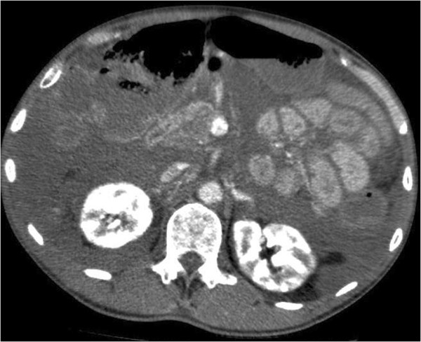
Figure 1.

CT Abdomen of patient 1, here at kidney–level, showing the dense mucous masses initially interpreted as pseudomyxoma peritonei filling the abdominal cavity, making the individual organs difficult to see. On this second CT-scan from day 2, the masses are increasingly dense, now measuring around 50 HU. There are no obvious signs of bowel ischemia.
