Figure 3.

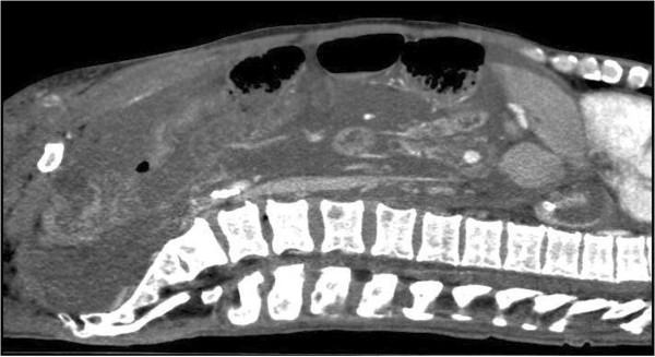
CT abdomen (patient 1), sagittal view with contrast, again illustrating what is seen as mucous masses filling the abdominal cavity, but which by ultrasonographic examination was revealed as massively thickened peritoneum.

CT abdomen (patient 1), sagittal view with contrast, again illustrating what is seen as mucous masses filling the abdominal cavity, but which by ultrasonographic examination was revealed as massively thickened peritoneum.