
Figure 3.
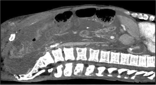
CT abdomen (patient 1), sagittal view with contrast, again illustrating what is seen as mucous masses filling the abdominal cavity, but which by ultrasonographic examination was revealed as massively thickened peritoneum.
Official websites use .gov
A
.gov website belongs to an official
government organization in the United States.
Secure .gov websites use HTTPS
A lock (
) or https:// means you've safely
connected to the .gov website. Share sensitive
information only on official, secure websites.
CT abdomen (patient 1), sagittal view with contrast, again illustrating what is seen as mucous masses filling the abdominal cavity, but which by ultrasonographic examination was revealed as massively thickened peritoneum.