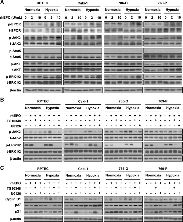
Figure 5.
Erythropoeitin activates the JAK and MAPK/ERK pathways. A, Four renal cell lines were starved in serum/growth factor-free media containing 0.1% BSA in normoxia or hypoxia. Cells were stimulated with the indicated concentration of rhEPO. Immunoblotting of protein extracts with indicated antibodies (at left of panel) shows JAK2 and MAPK/ERK pathway components in renal cell lines stimulated with rhEPO in the presence of hypoxia. β-actin is used as a loading control. B, Four renal cell lines were starved in serum/growth factor-free media containing 0.1% BSA in normoxia or hypoxia. Cells were subjected to 1 μM of TG10348 (a JAK2 inhibitor) or 1 μM of U0126 (a MEC inhibitor) for 60 mins prior to the addition of 10 units/mL rhEPO. Ten minutes after exposure of rhEPO, cell lysates were collected and subjected to Western blot analysis with the indicated antibodies. β-actin is used as a loading control. C, Cells are treated with the indicated concentrations of rhEPO in media containing 2% FBS for 24 hrs in normoxic or hypoxic condition. Cell lysates are subjected to Western blot analysis. Western blot analysis shows cyclin D1 was induced and p27 kip1 and p21 cip1 were down-regulated in renal cells stimulated with rhEPO in the presence of hypoxia. β-actin served as loading control.

