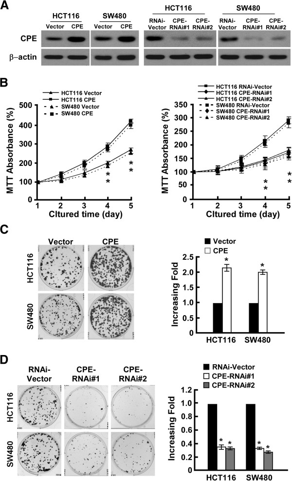
Figure 2.
CPE promotes colorectal cancer cell proliferation. (A) Western blot analysis of CPE expression in HCT116 and SW480 cell lines stably infected with CPE ORF or shRNA. β-actin was used as a loading control. (B) MTT assay analysis of cell growth rates for different stable cell lines at the indicated times after seeding cells. (C) Representative micrographs (left panel) and quantification (right panel) of colony formation in CPE-overexpressing and vector cells. (D) Representative micrographs (left panel) and quantification (right panel) of colony formation in CPE-silencing and vector cells. Data are presented as mean ± SD from three independent experiments. *: P < 0.05.

