Figure 5.
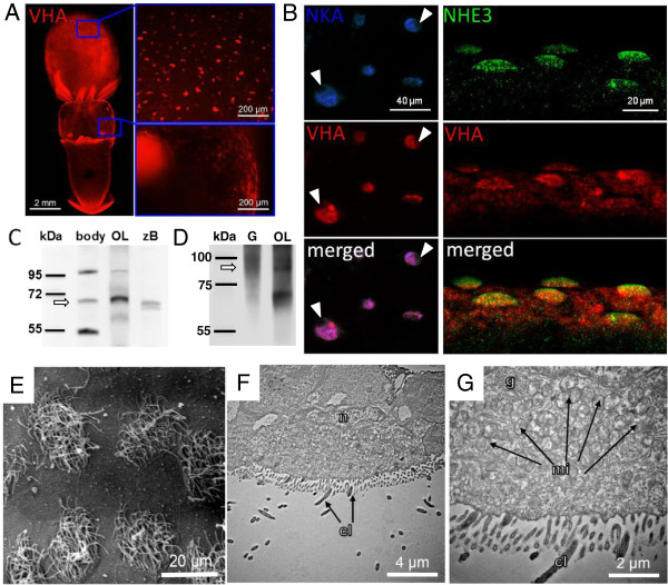
Immunohistochemical and electron microscopical analyses of epidermal ionocytes. Immunohistochemical detection of V-type H+-ATPase (VHA), Na+/K+-ATPase (NKA) and Na+/H+-exchanger 3 (NHE3) in epidermal ionocytes of squid Sepioteuthis lessoniana. VHA labeled ionocytes are distributed on yolk and head (overview image compiled from 11 images) (A). VHA labeled ionocytes are scattered in a “salt and pepper” pattern mainly on the ventral side of the yolk and head. Confocal images of double immuno-fluorescent labeling of NKA indicates co-localization with VHA positive cells (left panel) (B). VHA and NKA immuno reactivity demonstrates colocalization in epidermal ionocytes. Nuclei are covering the fluorescence signal (indicated by arrow head) suggesting a basolateral orientation of these transporters. Double labeling of VHA and NHE3 (right panel) confirms the basolateral orientation of VHA and demonstrates NHE3 localized in apical membranes (note the dome shaped apical surface of ionocytes labled by the NHE3 antibody). Western blot analysis of VHA subunit A in brain tissues of zebrafish (Danio rerio) (zB), optical lobe (OL), gills (G) and whole embryos (body) of squid (Sepioteuthis lessoniana), demonstrating immunoreactivity of the VHA antibody with a 70 kDa protein (C). Western blot analysis of NHE3 in brain, optical lobe (OL) and whole embryo (body) homogenates, demonstrating immunoreactivity of the NHE3 antibody with a 95 kDa protein (D) (bands indicated by arrows). Scanning and transmission electron microscopy of ciliated epidermal ionocytes of squid (S. lessoniana) embryos (E-G). Transmission electron microscopic images of surface epithelia demonstrating the ultrastuctural morphology of epidermal ionocytes in the head region which are particularly rich in mitochondria (F-G). cilia (cl), golgi (g), mitochondrion (mi), nucleus (n).

