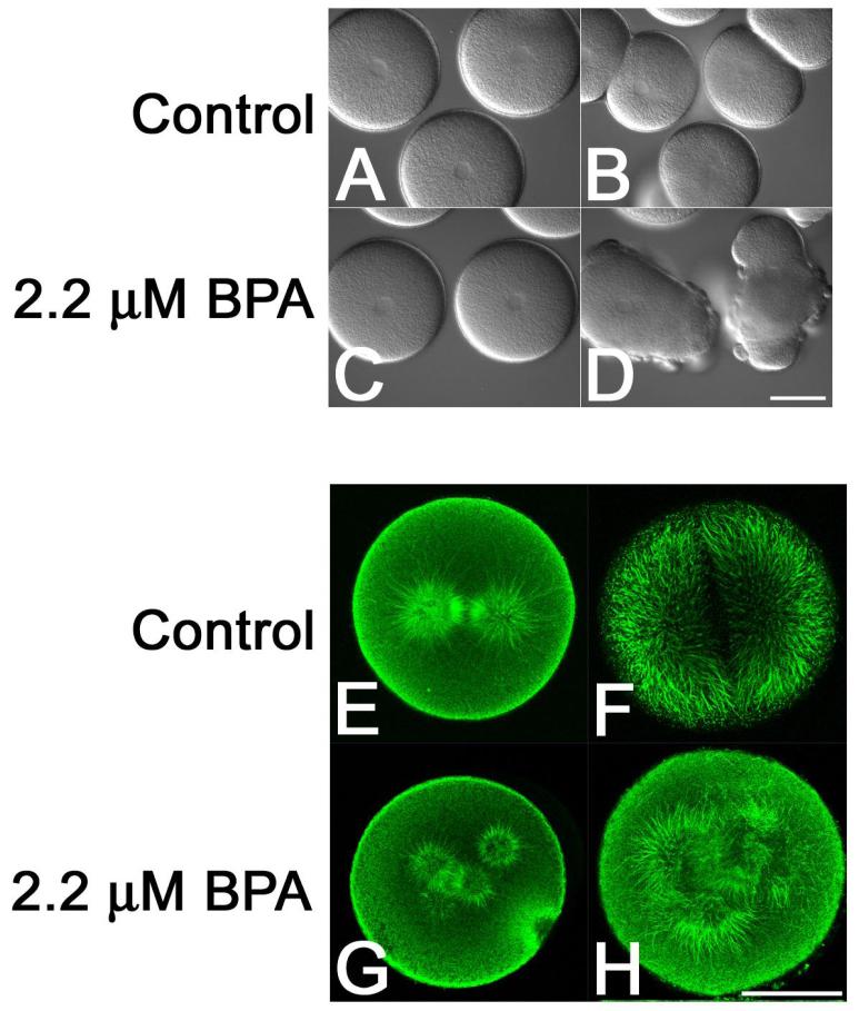
Figure 1. Dose-dependent effects of Bisphenol A on microtubule organization and cell division in sea urchin embryos.
L. pictus eggs were fertilized, stripped of their fertilization envelopes and cultured through the first division in the presence of O.1% DMSO (Panels A, B, E and F) or 2.2 μM BPA (Panels C, D G and H). Note that while the control embryos underwent normal cytokinesis (Panel B), BPA-treated embryos formed multiple, misplaced cleavage furrows (Panel D). Analysis of microtubule organization in metaphase (Panels E and G) and anaphase (F and H) embryos revealed the presence of normal metaphase and anaphase spindles in control embryos (Panels E and F) but supernumary spindle poles in BPA-treated embryos (Panels G and H). Bars for Panels D and H, 50 μm.

