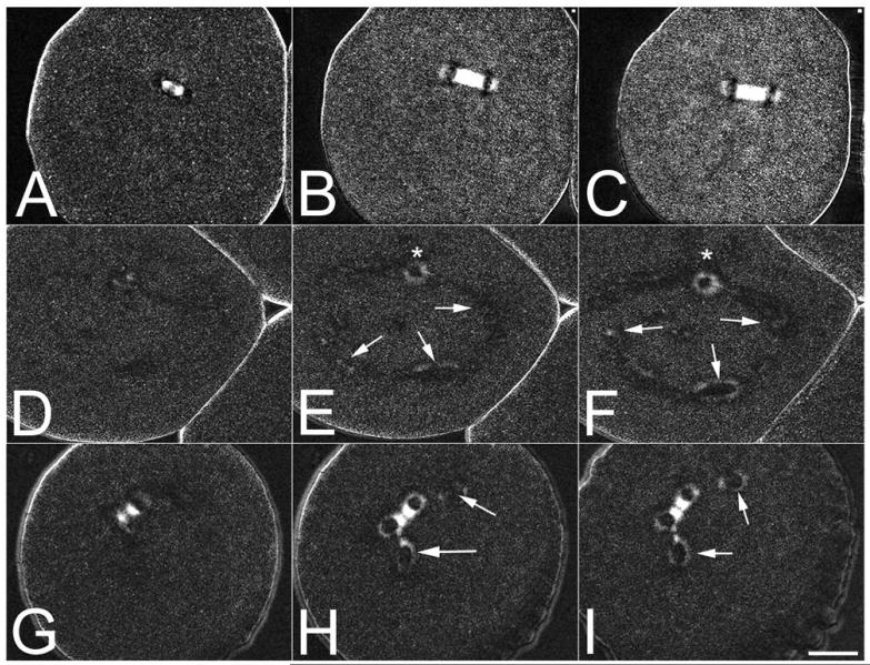
Figure 2. Visualization of ectopic spindle pole formation in Bisphenol A-treated sea urchin eggs.
L. pictus eggs exposed to either DMSO carrier (Panels A-C) or 2.2 μM BPA were compressed under fluorocarbon oil to aid in visualizing spindle formation, and followed by polarization microscopy. Whereas controls formed a normal, birefringent spindles (Panels A-C), embryos exposed to BPA could be observed forming monopolar spindles (*) as well as de novo microtubule organizing centers in the cytoplasm (Panels D-F, arrows). In other cells, asters could be observed splitting off the main spindle (Panels G-I, arrows). Bar 20 μM.

