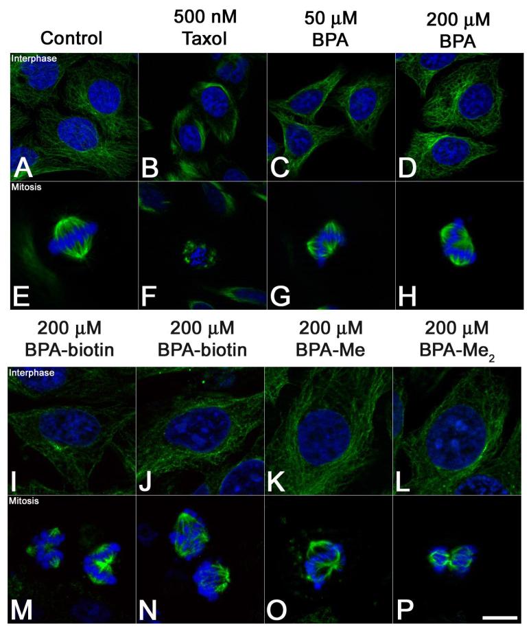
Figure 6. Ectopic spindle pole formation in HeLa cells exposed to Bisphenol A.
Hela cells were exposed to 0.1% DMSO (Panels A and E), 500 nM Taxol (B and F), or Bisphenol A (Panels C-D, F-H), BPA-biotin 1 (I and M), BPA-biotin 2 (J and N), Bisphenol A monomethyl ether (BPA-Me) (K and O), and Bisphenol A dimethyl ether (BPA-Me2) (L and P) for four hours, processed for DNA (blue) and tubulin (green) localization, and representative images were acquired from cells in interphase (Panels A-D; I-L) and mitosis (Panels E-H; M-P). In contrast to Taxol-treated cells (Panel B and F) that displayed small, stellate asters, BPA-treated cells developed ectopic spindle poles (Panels G and H). Similarly, biotinylated-BPA analogs produced phenotypes consistent with the parent molecule (Panels M and N), as did methylated BPA analogs BPA-Me and BPA-Me2 (Panels O and P). Bar 10 μm.

