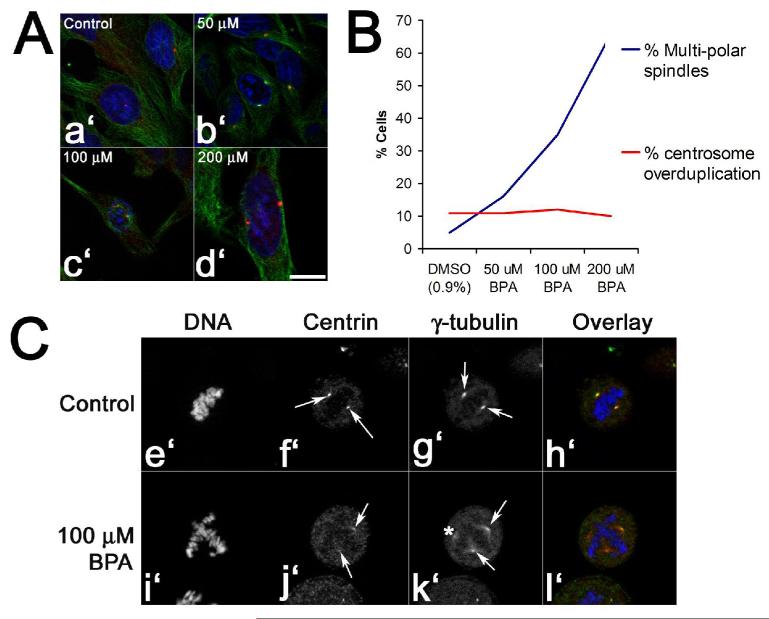
Figure 7. Bisphenol A does not drive centrosome amplification to generate ectopic spindle poles.
Panels A and B. Hela cells were exposed to either 0.1% DMSO or increasing doses of Bisphenol A for four hours, fixed and processed for DNA (blue), tubulin (green), and pericentrin (red) localization (Panel A, a’-d’; Bar, 10 μM). Cells were then scored for the presence of multipolar spindles or centrosomes at the G2/M transition (Panel B). Note that the number of pericentrin-positive centrosomes at G2/M does not increase even at 200 μM, where the majority of metaphase spindles are multipolar. Panel C. Centrin and γ-tubulin localization in control- (Panels e’ through h’) and BPA-treated eggs (Panels i’-l’). Compared to controls (Pictures f’ and g’, arrows), BPA-treated cells contained spindle poles that were positive for both γ-tubulin and centrin (Pictures j’ and k’, arrows), as well as diffuse poles that lacked centrin foci (asterisk k’). Bar, 10 μm.

