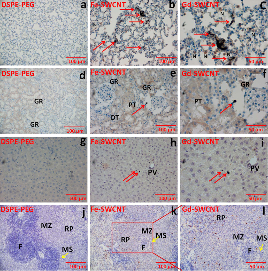
FIGURE 2.
Representative myeloperoxidase (MPO) immunostained specimens at day 1 of rat (a, b, c) lung (d, e, f), kidney (g, h, i), liver, and (j, k, l) spleen for PEG-DSPE, Fe-SWCNTs treatment, and Gd-SWCNT. Gd-SWCNT aggregates (red arrows) are found in lungs (b, c), kidney (e, f), liver (h, i), and spleen (k, l). The Gd-SWCNTs are aggregated in the alveolar epithelial lining of the lungs with few neutrophils (N) surrounding the Gd-SWCNTs; in the kidney glomerulus (GR) and in the hepatocytes of liver. The specimens do not show any signs of inflammation or tissue architectural damage. There were no changes in the MPO expression in lungs, kidney, liver, and spleen. GR, glomerulus; PT, proximal tubule; DT, distal tubule; PV, hepatic portal vein; F, follicle; MZ, marginal zone; MS, marginal sinus. [Color figure can be viewed in the online issue, which is available at www.interscience.wiley.com.]

