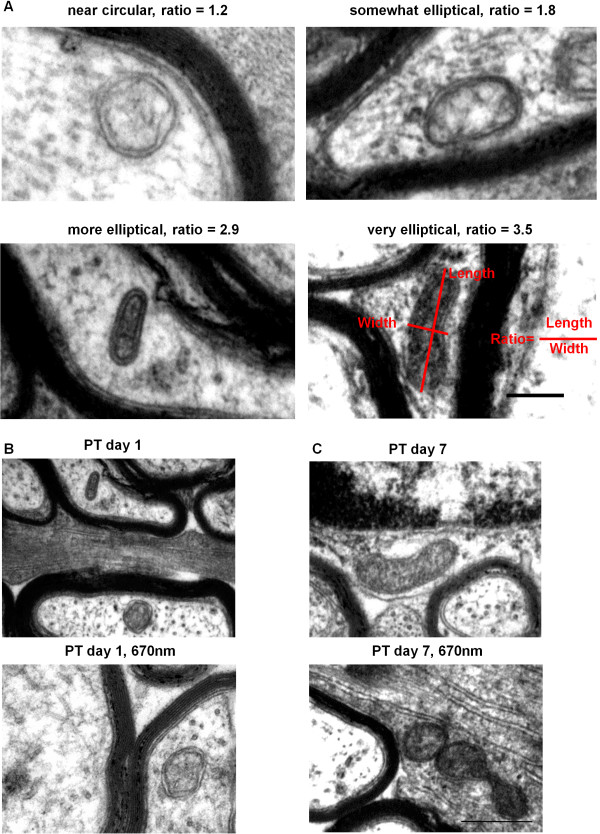
Figure 3.
Mitochondrial shape changes in secondary degeneration +/− R/NIR-IT. Representative images of shapes represented by common ratios, scale = 200 nm (A). Representative images illustrating lack of change in mitochondrial profile shape with R/NIR-IT at day 1 in axons (B) and more circular mitochondrial profiles with R/NIR-IT at day 7 in glia (C); scales = 500 nm.

