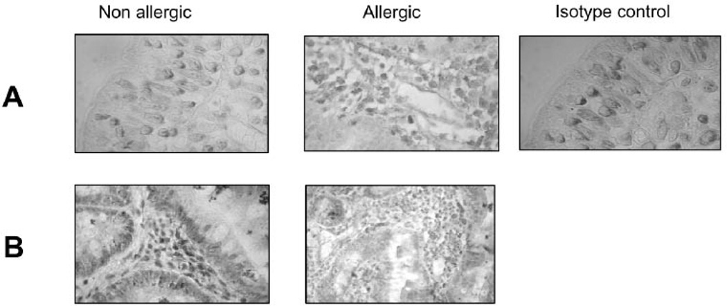
Fig. 2. Expression of Gal-1 and Gal-3 in duodenal specimens of allergic and non-allergic patients.
Gal-1 and Gal-3 expression was assessed by immuno-histochemistry using specific polyclonal antibodies against Gal-1 and Gal-3 (purified rabbit IgGs) in duodenal samples of allergic and non-allergic patients. Representative photographs of mucosa from allergic and non allergic patients were selected to show Gal-1 (A) and Gal-3 (B) positive cells. Control with serum from non-immunized rabbits is depicted. All samples were analyzed at least three times in independent experiments (Original magnification: 100x and 400x).

