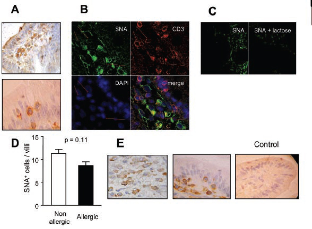
Fig. 3. SNA binding pattern on duodenal samples: Pattern of α2,6 sialylation in mucosal tissue of allergic versus non-allergic patients.
A) Lectin-histochemistry using biotinylated-SNA and hematoxylin staining was done to identify SAα2–6-galactose residues. A photograph with stained IEL from an allergic patient is shown. B) Co-localization studies using a Cy3-conjugated anti-CD3 antibody (red), biotinylated SNA/ FITC-conjugated streptavidin (green) and DAPI (blue) was performed by confocal microscopy. C) Lectin binding inhibition assessed by confocal microscopy. Cells were incubated with biotinylated SNA or biotinylated SNA in the presence of 100 mM lactose. D) SNA+ cells were quantified by lectin-histochemistry in biopsy specimens of allergic (N=8) and non-allergic (N=8) patients and no significant difference was found (p= 0.11). E) ST6Gal1 expression was detected using a polyclonal specific antibody by immunohistochemistry. A control of immunoreactivity is shown using sera from non-immunized rabbits. All samples were analyzed at least three times in independent experiments and representative images are shown (Original magnifications: 200x and 1000x).

