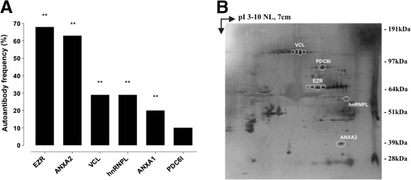
Figure 3.

Antigen validation in resectable PDAC patients. (A) The graph shows the frequency of autoantibodies against mouse and human common immunoreactive antigens in the group of resectable patients who underwent surgery with curative intent (n = 38), analyzed by SERPA against CF-PAC-1 cell line 2DE map. P-values were calculated vs. control frequencies listed in Table 3 by Fisher's exact test (** P < 0.005). (B) Proteins were extracted from eight frozen PDAC tissues from surgically-treated patients (stage IIA and IIB), separated by 2DE, transferred to a nitrocellulose membrane and probed with the autologous serum. A representative Western blot is shown; circles indicate the presence of autoantibodies against the mouse and human common immunoreactive antigens.
