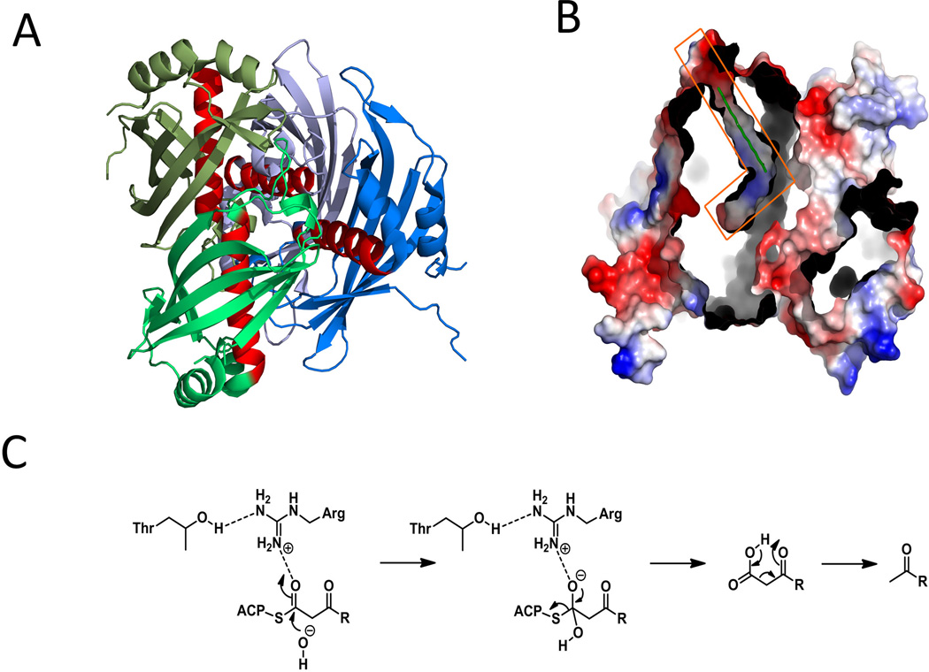
Figure 14.
Selected structures of hot-dog fold TE domains in PKS system. (A) hot-dog fold dynemicin TE DynE7 tetramer structures (“sausages” are in red). PDB code: 2XEM. (B) Surface representation of DynE7 tetramer and the L-shaped substrate binding channel in orange box. (C) Mechanism of decarboxylative hydrolysis in hot-dog fold TEs. 215×152mm (300 × 300 DPI).

