Figure 3.
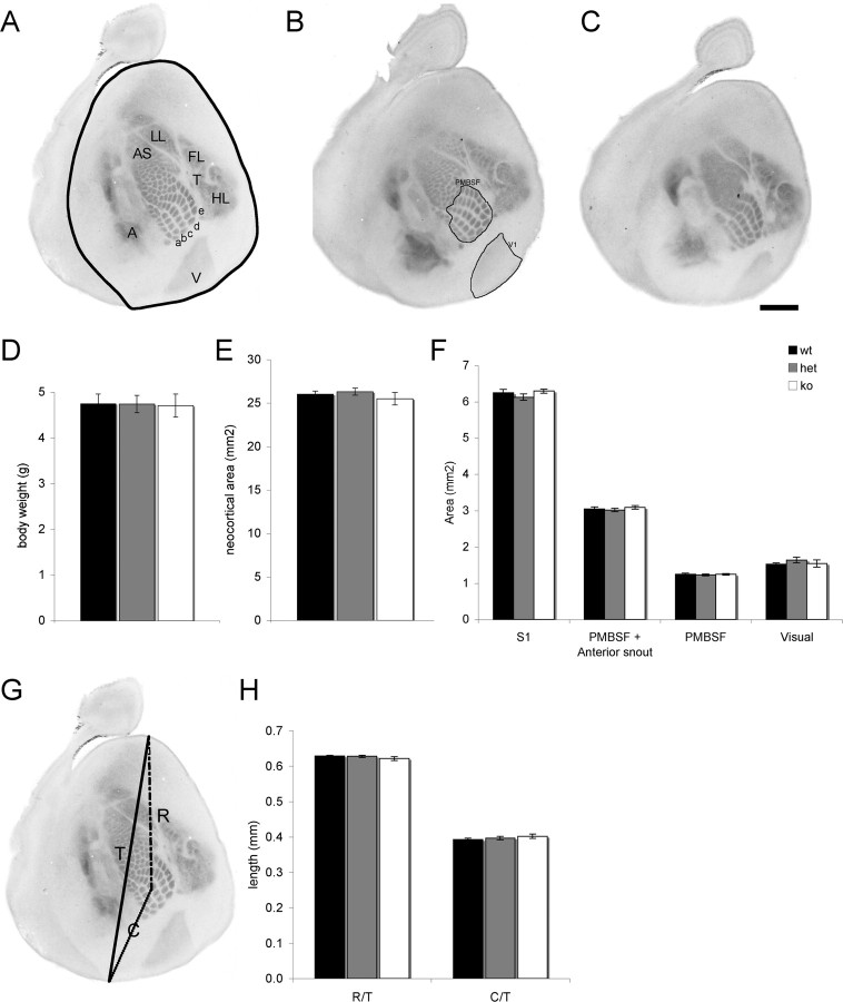
Loss of mGluR5 does not disrupt cortical arealization. Flattened sections through layer 4 of P7 Mglur5+/+ (n = 6) (A), Mglur5+/− (n = 9) (B), and Mglur5−/− (n = 9) (C) animals immunostained for 5-HTT to reveal thalamocortical afferents (TCA). The neocortex is outlined in A. TCA patch segregation appears to be normal in Mglur5+/− (B). However, in Mglur5−/− animals (C), 5-HTT staining is uniform in the anterior snout region (AS) indicating a lack of whisker-specific segregation. Although TCA patches are visible in PMBSF, their segregation appears reduced compared with Mglur5+/+ and Mglur5+/− animals. The primary visual (V), auditory (A), and somatosensory cortex (remaining regions) are clearly visible indicating that mGluR5 does not regulate TCA path finding. Body weight (D), neocortical area (E), and area of S1, PMBSF and visual cortex (F, boundaries for these areas are shown in A and B) are comparable among all genotypes. Similarly, the relative rostral (R)/caudal (C) position of S1 within the neocortical sheet is unaltered in Mglur5+/− and Mglur5−/− mice (G, H), indicating that the changes in pattern formation in mGluR5 mutant mice do not arise from a general delay in cortical development. Scale bar, 1 mm. wt = Mglur5+/+, het = Mglur5+/−, and ko = Mglur5−/−.

