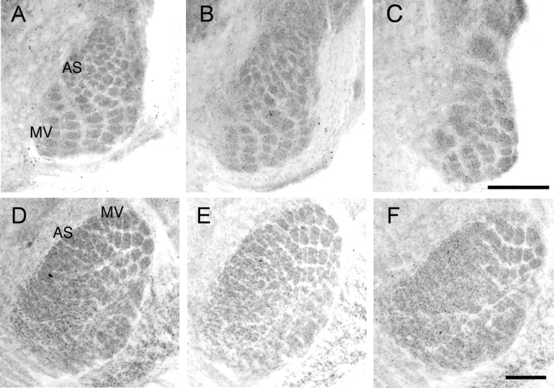
Figure 7.

Reduced barrelette and barreloid segregation in the anterior snout representation of PrV and VpM, respectively, at P7. Cytochrome oxidase staining in barrelettes in PrV (A–C) and barreloids in VpM (D–F) from Mglur5+/+ (A, D), Mglur5+/− (B, E), and Mglur5−/− (C, F). In Mglur5−/− mice, whisker-related patches are clearly visible in regions receiving input from the main mystacial vibrissae (MV). In contrast, in the region of PrV that receives input from the anterior snout whiskers (AS), the barrelette pattern is clearly distorted and no segregation is seen in the corresponding region in VpM. Scale bars: C (for A–C), F (for D–F), 250 μm.
