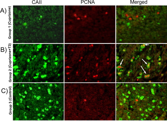
Figure 8.
A–C, Double immunostaining (CA II and PCNA) of the corpus callosum of mice from group 1 (A), group 2 (B), and group 3 (C). Numerous cells double stained with PCNA and CA II markers were present in the corpus callosum of T3-treated mice (B). The double-labeled cells were absent from the corpus callosum of chronically demyelinated mice (A) or control mice (C). Magnification ×600.

