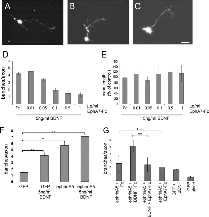
Figure 1.
EphA7–Fc abolishes in a concentration-dependent manner BDNF-promoted branching of retinal axons. Single cells were prepared from nasal thirds of E8 chick retinas, electroporated with an eGFP expression plasmid to facilitate a later analysis of neurite outgrowth and branching, and plated at low density on laminin/merosin-coated dishes. After 4 d in culture, typical branching patterns have emerged. A, Little branching without BDNF. B, Strong branching in the presence of 5 ng/ml BDNF and 1 μg/ml Fc. C, Strong reduction in branching after application of 5 ng/ml BDNF and 0.5 μg/ml EphA7–Fc. Scale bar, 10 μm. D, A quantification of branching from three independently performed experiments with each n > 15. E, The lengths of the axons in various EphA7–Fc concentrations were not affected. Statistical analysis was performed using ANOVA. F, Overexpression of ephrinA5 on retinal axons results in an increase in branching, which can be further enhanced by treatment with BDNF. Statistical analysis was performed using ANOVA with Dunnett's post hoc test. G, The increase in branching caused by BDNF, on top of a higher branching caused by a moderate overexpression of ephrinA5, can be abolished by treatment with EphA7–Fc (1 μg/ml), whereas treatment with EphA7–Fc in the absence of BDNF has no effect on branching. Analyses were done by one-way ANOVA and Tukey's multiple-comparison test.

