Figure 7.

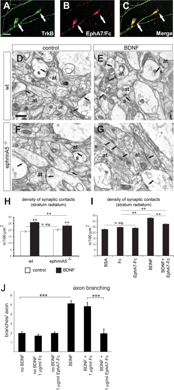
EphrinA5 cooperates in BDNF-induced synaptogenesis in hippocampal neurons. A–C, Double-labeling confocal images showing two examples of axonal growth cones (arrows) from hippocampal explants after 4 d in culture. Axonal growth cones coexpress TrkB (viewed by Alexa488-labeled antibody; A) and ephrinAs (using EphA7–Fc and viewed by Texas Red; B) as displayed in merged images (C). D–G, Electron micrographs illustrating axon terminals and synaptic contacts (arrows) in the stratum radiatum of organotypic cultures from wild-type and ephrinA5−/− mice under control conditions (D and F, respectively) and after treatment with BDNF (E and G, respectively). H, Density of synaptic contacts in the stratum radiatum of control and treated organotypic cultures from ephrinA5−/− mice and their littermates. I, Histogram showing density of synaptic contacts in the stratum radiatum of 13 d in vitro organotypic cultures under control conditions (BSA) or treatment with Fc, EphA7–Fc, recombinant human BDNF (BDNF), and recombinant human BDNF plus EphA7–Fc (BDNF+EphA7–Fc). Mean ± SEM; n.sig, nonsignificant; **p < 0.01, Student's t test; ***p = 0.001. at, Axon terminal; s, dendritic spine. Scale bars: A–C, 10 μm; D–G, 0.5 μm. J, EphA7–Fc treatment abolishes the BDNF-induced branching of hippocampal neurons. Using a protocol described in experiments documented in Figure 1, hippocampal neurons were treated for 2 d under various conditions as indicated.
