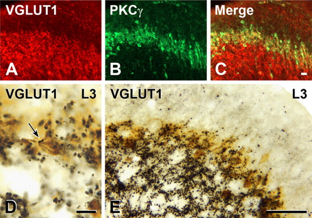
Figure 3.
Myelinated primary afferents that express VGLUT1 (in the rat) terminate among PKCγ interneurons. A–C, VGLUT1-immunoreactive axons occur in the layer containing PKCγ-immunoreactive interneurons (yellow; C). D, Two VGLUT1-immunoreactive terminals (arrow) form close appositions on the cell body of a PKCγ-immunoreactive neuron (brown) that lies within the dense band of PKCγ neurons in lamina IIi (taken from the L3 segment). E, In segment L3 as in other spinal segments, the dense band of PKCγ neurons in lamina IIi is heavily innervated by VGLUT1-immunoreactive axons. Scale bars: A–C (in C), D, 10 μm; D, 10 μm; E, 50 μm.

