
Figure 2.
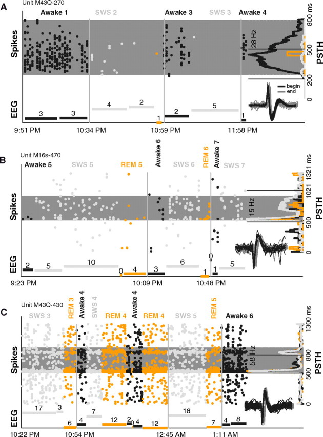
Example units modulated during sleep. A, Spiking activity and EEG measured during different time points while the animal slept (data were not collected continuously). This unit's response to a sAM tone (carrier frequency = 8.3 kHz, modulation frequency = 256 Hz, sound level = 30 dB SPL) consistently went down when the unit was tested in sleep (gain = −96% in SWS) (dark gray, awake; light gray, SWS; and orange, REM). The spike raster (oriented vertically, each column represents a single trial) shows a clear loss of spiking on entry into SWS in episode 2, a recovery of response on returning to awake in episode 3, and a loss of response again in SWS of episode 4. Far right, The PSTH averages the activity across all trials for each state and is plotted on its side. The clear response in awake is absent in SWS and REM. Gray shaded strip is the analysis window determined by our windowing algorithm. Dashed lines demarcate stimulus playing. Bottom, The normalized EEG amplitude (minimum subtracted) for each epoch tested is shown (times each episode began are marked on abscissa; breaks between EEG bars within an episode denote noncontinuous measurements; and numbers above each bar denote duration of measurement in minutes). When driven spiking is high in awake periods, EEG amplitude is low. In SWS periods, high EEG amplitude accompanies diminished firing. Inset, first 50 (black) and last 50 (gray) spike waveforms collected of the unit during the 2.5-h-long session. Horizontal offset is for display purposes only. B, Same format as A. This unit's response to a marmoset vocalization sound (type = tsik, sound level = 80 dB SPL) went up in SWS (gain = 86% in SWS) but not in REM. Firing rate increased when EEG amplitude (SWS) increased. Session spanned 2 h. C, Same format as A. Unit's response to a tone (frequency = 8.7 kHz, sound level = 60 dB) was similar in awake, SWS, and REM (gain = −11% in SWS). Regardless of multiple large changes in EEG amplitude and behavioral state over the 3-h-long session, the unit remained consistently well driven by the stimulus, and the PSTH retained its distinct onset and offset pattern in all three states.