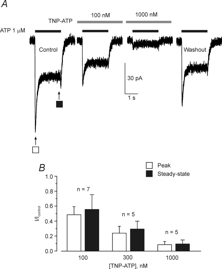
Figure 5.
Inhibition of ATP-induced currents by TNP-ATP. A, ATP-induced current measured for the cortical astrocytes in control conditions, in the presence of TNP-ATP, and after the washout. B, Mean data showing the percentage of ATP-induced current inhibition at three different concentrations of TNP-ATP; the number of experiments is shown on the graph. Application of TNP-ATP started 2 min before application of ATP. All recordings were made at a holding potential of −80 mV.

