
Figure 5.
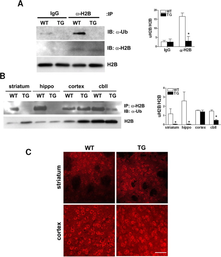
uH2B is decreased in transgenic R6/2 brain. A, Western blot showing that the global uH2B level in 8 week transgenic R6/2 whole brains was below a detection limit of immunoprecipitation (IP) with IgG or anti-H2B antibody followed by immunoblotting with anti-ubiquitin antibody (IB: α-Ub). This blot was stripped and reprobed with anti-H2B antibody, which showed that the same amounts of H2B were pulled down from wild-type and transgenic mice (IB: α-H2B). Western blot analysis of histone extracts also demonstrated H2B expression levels were equal in wild-type and transgenic mice (H2B). Densitometry of uH2B/H2B is shown on the right (n = 4 animals per group; *p < 0.05). B, Western blot showing global uH2B levels are undetectable in 4 week transgenic striatum and hippocampus and low in 4 week transgenic cerebellum. Western blot analysis of histone extracts from each brain region of 4 week mice demonstrated equal expression levels of H2B in wild-type and transgenic mice (H2B). Densitometry of uH2B/H2B is shown on the right (n = 4 animals per group; *p < 0.05). C, Confocal images of 12 week wild-type and transgenic brains stained with anti-hBre1 serum (scale bar, 50 μm). Error bars indicate SEM. TG, Transgenic; WT, wild type; hippo, hippocampus; cbll, cerebellum.