
Figure 2.
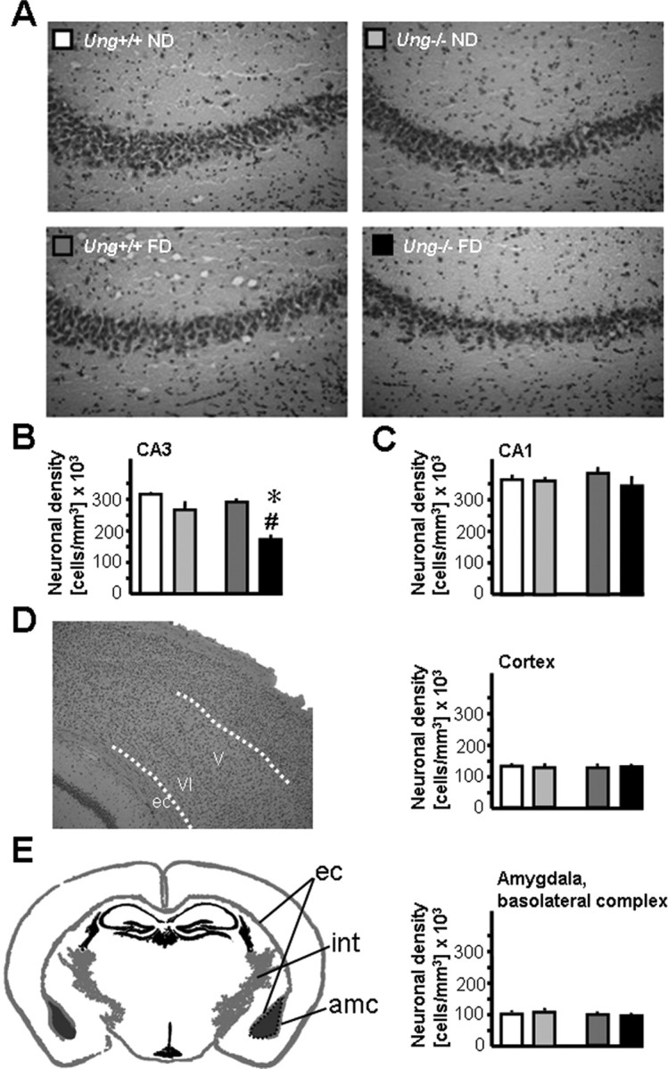
Increased susceptibility of Ung−/− mice to neurodegeneration after chronic folate depletion in vivo. A, Representative images of 10-μm-thick H&E-stained sections illustrating reduced neuronal density in CA3 of Ung−/− animals as a consequence of chronic folate deficiency (bottom right). B, Density of CA3 pyramidal neurons; ANOVA for genotype: F (1,16) = 22.8, p < 0.0005; treatment: F (1,16) = 11.3, p < 0.005; interaction: F (1,16) = 3.8, p = 0.068; 1-β = 0.44. C–E, Similar neuronal densities in other brain regions. C, Hippocampal subfield CA1. D, Deep cortical layers of frontoparietal cortex. E, Basolateral complex of amygdala. ec, External capsule; int, internal capsule; amc, amygdalar capsule. *p < 0.05, ND versus FD; # p < 0.05, Ung+/+ versus Ung−/−.