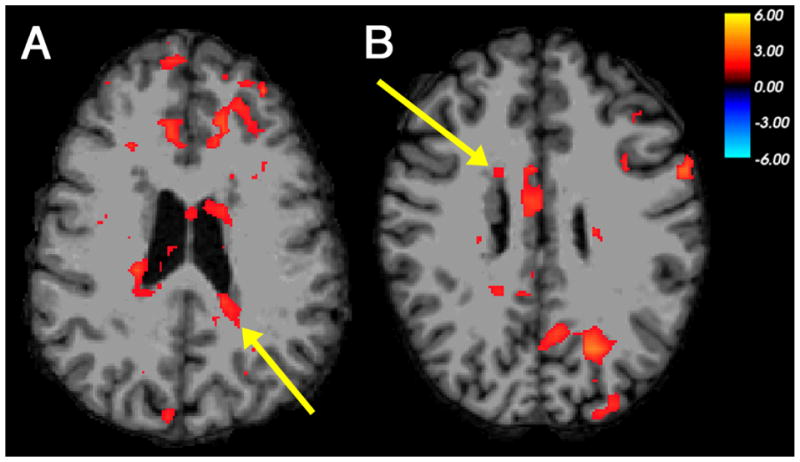
Figure 1. Co-activation of nodular heterotopia with multiple cortical regions in phonological processing.

Examples of BOLD activation seen in periventricular nodules of gray matter and multiple regions of cerebral cortex, in a functional contrast between picture-mediated word rhyming and word matching judgments as performed by individuals with PNH. (A) Left posterior heterotopia activation (arrow), with co-activation in bilateral frontal cortex, in Participant 2. (B) Right frontal heterotopia activation (arrow), with co-activation in contralateral posterior cortex, in Participant 4. The color scale represents the t-statistic for a significant contrast at each voxel. The right side of the images represents the left side of the brains.
