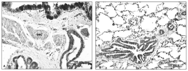
FIGURE 5.

Intense staining for iNOS in sheep lung 4 hours after S+B injury. (A) Micrograph depicting increased staining of the entire bronchial epithelium (E), mucous glands (MG), and smooth muscle cells. (B) After injury, increased iNOS expression is also present in bronchiolar epithelium (E), endothelial cells (EC), and alveolar macrophages (M) in the parenchyma.
