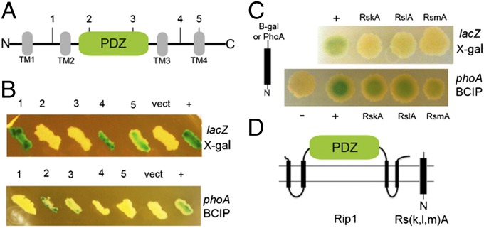
Fig. 1.
Membrane topology of Rip1 and its three anti-sigma factor substrates. (A) Schematic representation of M. tuberculosis Rip1 illustrating the four predicted transmembrane domains (TM1 through -4, in gray) and the PDZ domain between TM2 and 3 (in green). The vertical lines marked with numbers 1–5 mark the fusion points to either β-galactosidase or alkaline phosphatase as follows: 1, Y89; 2, V145; 3, W218; 4, G361; and 5, L383. (B) Membrane topology of Rip1. lacZ (Upper) and phoA (Lower) fusions to Rip1 at the positions indicated in A were expressed in M. smegmatis along with a vector control (vect) or a positive control (+) for lacZ (consisting of unfused lacZ) or phoA [an antigen85-phoA fusion (26)]. The upper image shows cells bearing each β-galactosidase fusion cultured on media containing X-gal and the bottom panel shows each PhoA fusion cultured on media containing BCIP. (C) Membrane topology of Rip1 substrates. β-Gal or PhoA was fused to the C terminus of Anti-SigK (RskA), Anti-SigL (RslA), or Anti-SigM (RsmA), and the plasmids encoding these fusions were transformed into M. smegmatis and cultured on media containing X-gal (Upper) or BCIP (Lower). The positive controls for X-gal and BCIP are the same as used in B. (D) Experimentally determined topology of Rip1 and its substrates.

