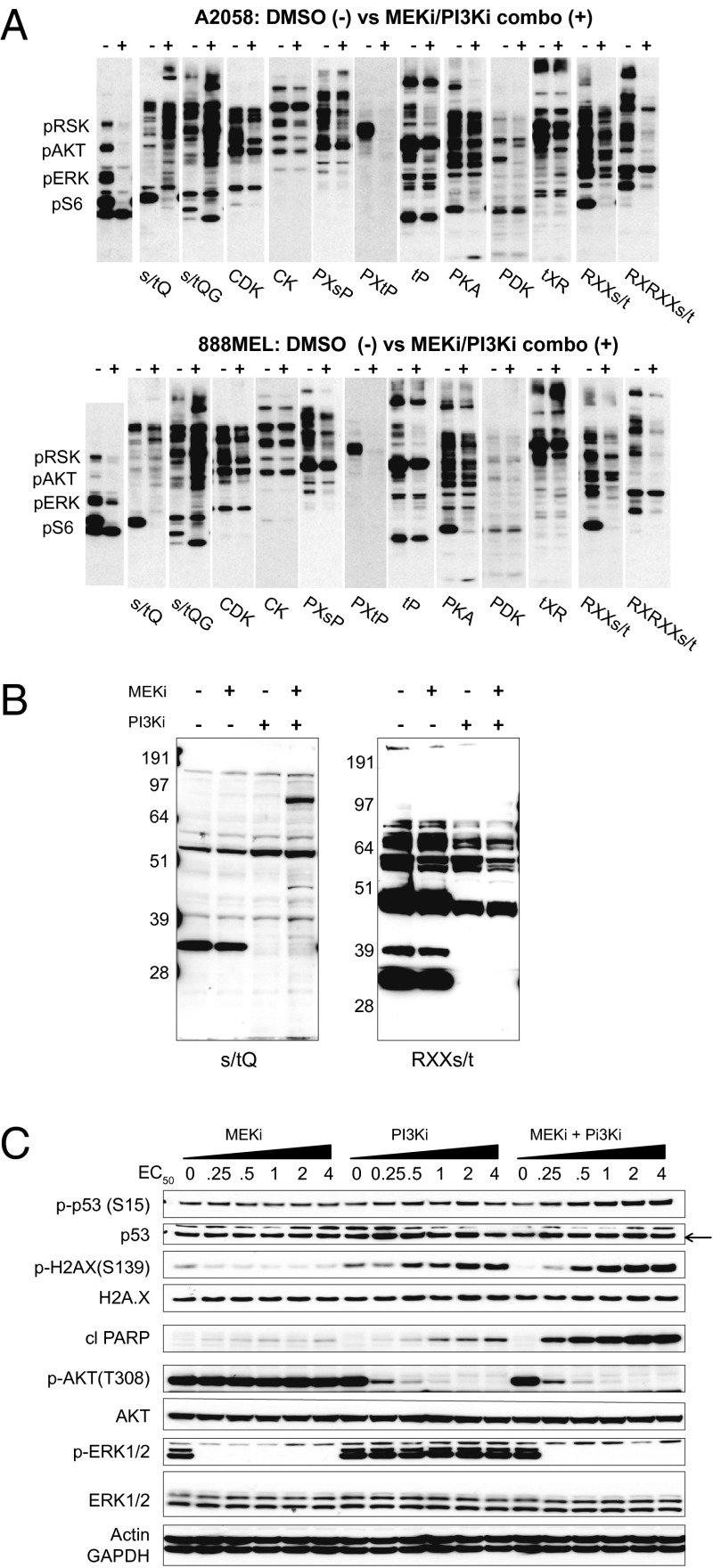
Fig. 1.
Dual inhibition of MEK and PI3K induces phosphorylation of DDR substrates. (A) A2058 or 888MEL melanoma cells were treated for 6 h with DMSO or GDC-0973+GDC-0941 (MEKi/PI3Ki combo; 4× EC50) and were subjected to KinomeView Profiling. A2058 and 888MEL cells were treated with 10 μM GDC-0973 + 10 μM GDC-0941 or 0.2 µM GDC-0973 + 10 µM GDC-0941, respectively. Blots were probed using an antibody mixture recognizing pRSK, pAKT, pERK, and pS6 or phosphomotif antibodies (e.g., DDR substrates with the [s/t]Q motif and AKT substrates with the RXX[s/t] and RXRXX[s/t] motifs). (B) A2058 lysates probed with the [s/t]Q and RXX[s/t] antibodies after treatment with DMSO, 10 μM GDC-0973 (MEKi), 10 μM GDC-0941 (PI3Ki), or the combination. (C) Dose response of A2058 cells to increasing concentrations of MEKi and PI3Ki alone or in combination. Blots were performed against DDR (p53 pSer15, histone 2AX pSer139), cell survival/cell death (AKT pThr308, cleaved PARP), and cell signaling (ERK1/2 pThr202/Tyr204) markers and controls. Actin and GAPDH served as loading controls.

