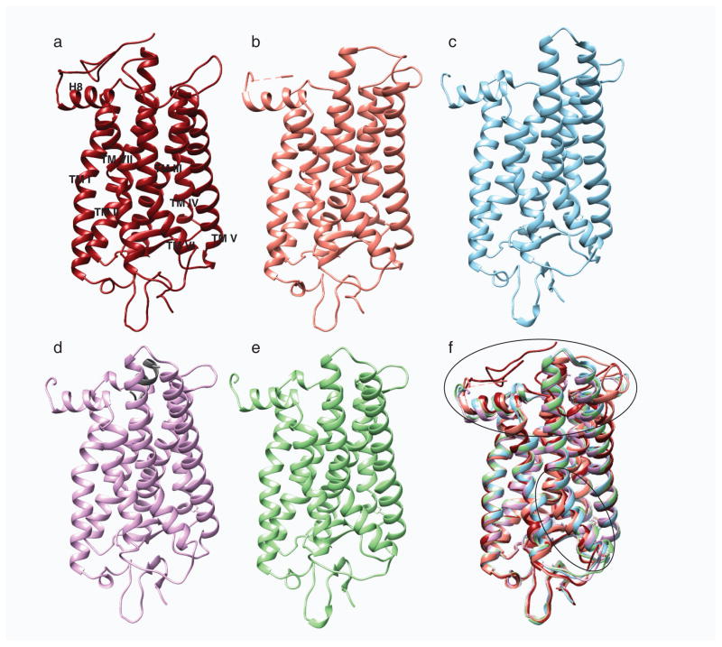
Fig. 2.
Structural comparison of different photo-states of rhodopsin. a, Structures of ground state rhodopsin (1u19) (Pescitelli et al. 2008); b, photoactivated rhodopsin (2i35) (Salom et al. 2006); c, opsin (3cap) (Park et al. 2008); d, opsin with bound C-terminal peptide of Gt (3dqb) (Scheerer et al. 2008) and e, Meta II (3pxo) (Choe et al. 2011) are shown in dark red, salmon, blue, purple and green, respectively. f, All activated rhodopsin states were superposed on its ground state structure. Superposition of these structures show rearrangement of the cytoplasmic loops and movement of helix TM VI depicted with black ovals.

