
Figure 5.
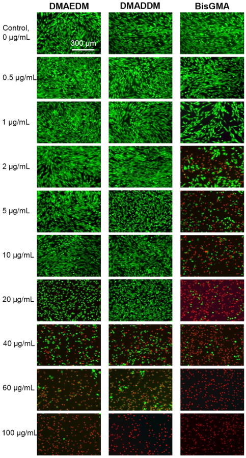
Representative live/dead staining images of human gingival fibroblasts (HGF) cultured in medium containing monomers. The top titles indicate the monomer names. The left side labels indicate the monomer concentrations in the culture medium. Live cells were stained green, and dead cells were stained red. BisGMA showed the most rapid decrease in green cells with a higher proportion of red cells when the monomer concentration was increased.