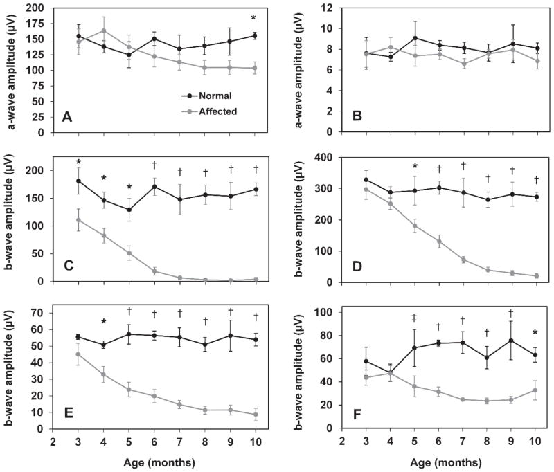
Figure 2.
ERG amplitudes as a function of age in normal and CLN2-affected dogs. ERG a-wave amplitude from 10 cd.s/m2 scotopic (A), and 3 cd.s/m2 photopic (B) recordings. Significant deficits in a-wave amplitude are not present until 10 months of age and only with scotopic recordings. ERG b-wave amplitude from scotopic rod responses (C), scotopic mixed responses from rods and cones at 10 cd.s/m2 (D), and photopic cone (E) and 30 Hz flicker recordings (F). Significant differences in b-wave amplitude exist even in early stages of disease (†, p<0.001; *, p<0.005; ‡, p<0.05). Error bars represent SEM.

