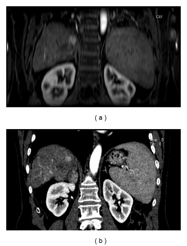
Figure 1.

77-year-old man treated with RFA for HCC. (a) Arterial-phase, coronal Vibe T1-W FS at 1-month followup. Residual tumor tissue in the cephalad portion of the treated area. (b) Coronal reformatted, artery-phase CT at 1-month followup. Residual tumor tissue in the cephalad portion of the treated area.
