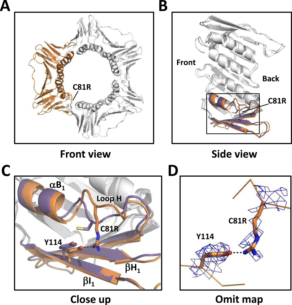
Figure 2.
Structure of the C81R mutant PCNA protein. (A) Front view of the C81R mutant PCNA protein trimer with one of the subunits shown in orange. (B) Side view of one subunit of the C81R mutant PCNA protein with the α-helix B1 and the β-strands H1 and I1 shown in orange. The wild-type PCNA structure is overlaid with the same α-helix and β-strands shown in purple. (C) Close up of the region near the C81R substitution. The mutant PCNA protein is shown in orange and the wild-type PCNA protein is shown in purple. (D) 2Fo-Fc omit map contoured at 1 σ and carved at 1.7 Å around the substituted arginine at residue 81 and Tyr-114.

