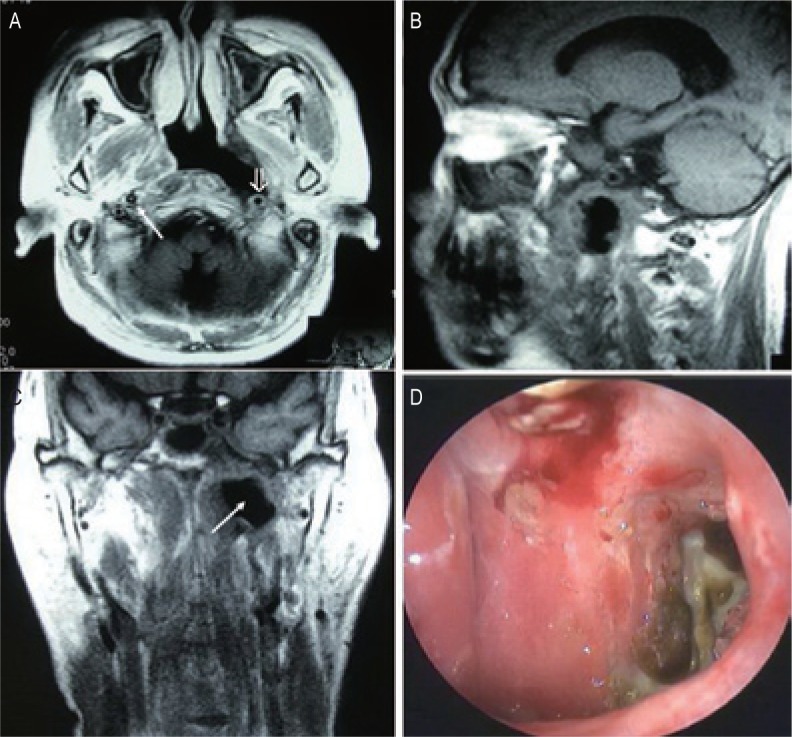
Figure 2. MRI and endoscopic findings of a postradiation nasopharyngeal necrosis with erosion and carotid exposure.

MRI images were obtained on a 56-year-old man who underwent CRT after the second course of radiotherapy. The man died of massive bleeding 3 months after the diagnosis of postradiation nasopharyngeal necrosis. A, transverse T1-weighted image showing nasopharyngeal necrosis is mainly located in the left wall of nasopharyngeal cavity. There is a defect in the left parapharyngeal space, and the internal carotid artery is involved (open arrow). Note the normal right carotid artery for comparison (arrow). B, sagittal T1-weighted image showing a defect in the posterior wall and the discontinuity of the posterior soft tissue. C, coronal T1-weighted image showing the defect in the left wall is extended to the left parapharyngeal space (arrow). D, necrosis is located in the left wall of the nasopharyngeal cavity, and the cavity is covered with secretion and necrotic tissue (observed under telescope 0°).
