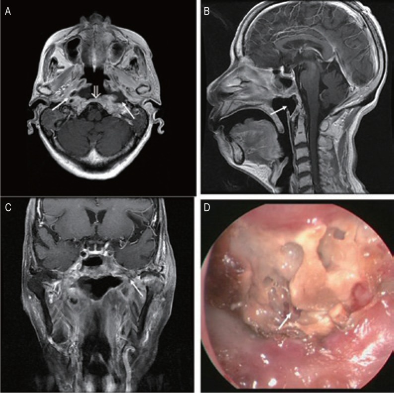
Figure 3. MRI and endoscopic findings of a postradiation nasopharyngeal necrosis with extension erosion and osteoradionecrosis.

MRI images were obtained on a 74-year-old woman who underwent CRT. A, transverse T1-weighted image shows that the destruction of the bone was extensive and that the lesions involved the clivus (open arrow) and bilateral parapharyngeal space (arrows). B, sagittal T1-weighted image showing a hollow defect in the posterior wall (arrows). C, coronal T1-weighted image shows that the necrosis is extensive and small air bubbles are present (arrow). D, the necrosis is located in the posterior wall of the nasopharyngeal cavity and the sequestra can be seen within the necrotic bones (arrow; observed under telescope 0°).
