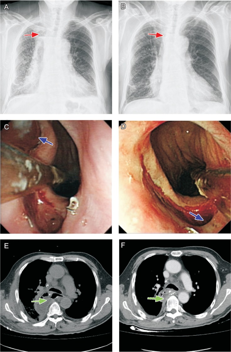
Figure 2. The results of clinical examinations before and after treatment.

A, chest X-ray radiography shows the gas-fluid level (red arrow) in the right superior mediastinum caused by the fistulae. B, the encapsulated effusion disappeared after transluminal drainage (red arrow). C, endoscopy shows that a transluminal drainage tube (blue arrow) was inserted into the fistulae for irrigation and section. D, the fistulae was healed after removal of the drainage tube (blue arrow). E, chest computed tomography shows partial abscess (green arrow) in the right chest before transluminal drainage. F, the abscess decreased markedly after transluminal drainage (green arrow).
