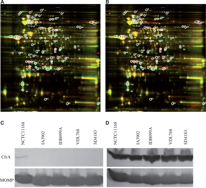
Fig. 4.—
Comparative proteomics analysis of Campylobacter jejuni strains by 2D-DIGE and Western blotting analysis of CfrA expression. (A) Overlay image for in-gel comparison between IA3902 (red) and NCTC11168 (green). (B) Overlay image for in-gel comparison between IA5908 (red) and NCTC11168 (green). The circled and numbered protein spots in gel A and gel B indicate the differentially expressed protein spots between the strains, and those in gel A were extracted and subjected to MALDI TOF/TOF mass spectroscopy. (C) Western blotting analysis of the whole cell lysates of C. jejuni isolates grown in MH broth. (D) Western blotting analysis of the whole cell lysates of C. jejuni isolates grown in MH broth supplemented with desferrioxamine (20 µM). The CfrA protein was detected with an anti-CfrA antibody, while MOMP, which is used as an internal control, was detected with a MOMP-specific antibody.

