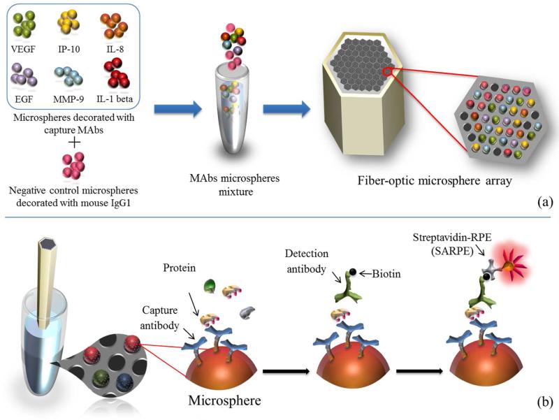
Figure 1.
Workflow of the multiplexed protein microarray. (a) Microarray assembly process: seven different types of microspheres (including one negative control) were mixed and loaded into the microwells on the etched fiber-optic bundle. (b) Saliva analysis: the assembled microarray was incubated sequentially with the saliva sample, a mixture of different biotinylated detection antibodies, and SARPE dye.

