
Figure 8.
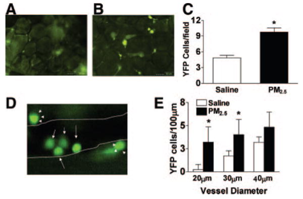
PM2.5 exposure increases YFP cell infiltration in mesenteric tissue and YFP cell adhesion in the cremasteric vasculature in c-fmsYFP mice fed HFC. A, B, Representative images of YFP cells in mesenteric tissue treated by either saline (A) or PM2.5 (B) and the quantification of YFP cells (C). D, Representative image of YFP cells in cremasteric tissue treated by PM2.5. The arrows depict YFP cells inside the vessel; closed arrowheads, YFP cells outside the vessel. E, Quantification of adherent YFP cells. The postcapillary venule boundaries are outlined in white lines. n=4. *P<0.05, saline vs PM2.5.