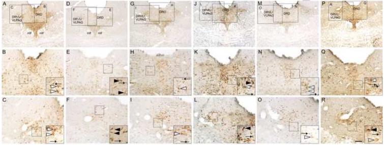
Figure 6.
Photomicrographs illustrating c-Fos/tryptophan hydroxylase (Tph) immunostaining in the mid-rostrocaudal dorsal raphe nucleus (DR; −8.00 mm bregma) in representative rats from each treatment group. Photomicrographs illustrate immunostaining in rats exposed to (A-C) HC/HC, (D-F) HC/25 °C, (G-I) 19 °C/HC, (J-L) 19 °C/25 °C, (M-O) 25 °C/HC, (P-R) 25 °C/25 °C. Black boxes in A, D, G, J, M and P represent regions shown at higher magnification in B, C, E, F, H, I, K, L, N, O, Q and R. Black boxes in B, C, E, F, H, I, K, L, N, O, Q and R indicate regions shown at higher magnification as insets located in the lower right-hand corner of these respective panels. Black arrows indicate representative examples of c-Fos-immunoreactive (ir) non-serotonergic cells (blue/black nuclear staining); white arrowheads indicate representative examples of Tph-ir/c-Fos-immunonegative neurons (red/brown cytoplasmic staining); black arrowheads indicate representative examples of c-Fos-ir/Tph-ir neurons (red/brown cytoplasmic staining with blue/black nuclear staining). Scale bar = 250 μm (A, D, G, J, M, and P); 100 μm (B, C, E, F, H, I, K, L, N, O, Q, and R); 50 μm (insets).

