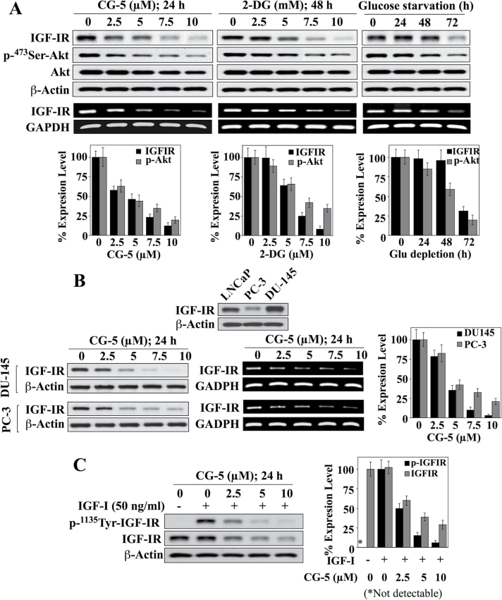
Fig. 1.
Effects of the glycolysis inhibitors CG-5, 2-DG and glucose-depleted medium on IGF-IR expression in prostate cancer cells. (A) Upper panels, western blot and/or RT–PCR analyses of concentration-/time-dependent effects of CG-5, 2-DG and glucose deprivation on IGF-IR expression and Ser473-Akt phosphorylation in LNCaP cells in 10% FBS-supplemented RPMI 1640 medium. Lower panels, relative expression levels of IGF-IR protein and p-Ser473-Akt. Amounts of immunoblotted proteins were analyzed by densitometry and expressed as a percentage of that in the respective control group (n = 3). (B) CG-5-mediated inhibition of IGF-IR expression in DU-145 and PC-3 cells. Upper panel, differential protein expression levels of IGF-IR in three prostate cancer cell lines, LNCaP, PC-3 and DU-145. Lower left and middle panels, western blot and RT–PCR analyses of the concentration-dependent effect of CG-5 on the protein and mRNA expression levels of IGF-IR in DU-145 and PC-3 cells. Lower right panel, relative protein expression levels of IGF-IR in response to CG-5 treatment in DU-145 and PC-3 cells. Protein abundance was analyzed by densitometry and expressed as a percentage of that in the control group (n = 3). (C) Western blot analysis of the concentration-dependent suppressive effects of CG-5 on the phosphorylation and expression levels of IGF-IR in LNCaP cells cotreated with IGF-I or vehicle control (left) and the corresponding densitometric analysis of relative expression levels (right; n = 3). All western blots shown are representative of three independent experiments. *P < 0.05.

