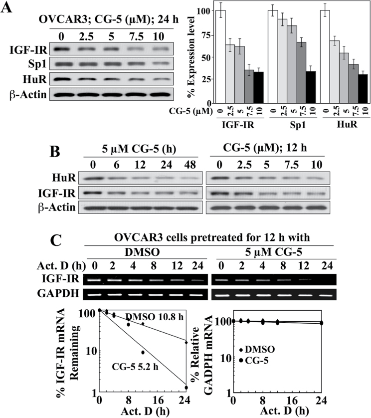
Fig. 4.
HuR acts as a stabilizing protein for IGF-IR mRNA in OVCAR-3 ovarian cancer cells: effects of CG-5 on protein expression and mRNA stability. (A) Representative immunoblot of the concentration-dependent effects of CG-5 on the expression of IGF-IR, Sp1 and HuR in 10% FBS-supplemented RPMI 1640 medium after 24 h of treatment (left) and the corresponding densitometric analyses of relative expression levels (right; n = 3). (B) Western blot analyses of the time-dependent effect of 5 µM CG-5 (left) and the concentration-dependent effect of CG-5 after 12h of treatment (right) on the expression of HuR versus IGF-IR in 10% FBS-supplemented RPMI 1640 medium. (C) Upper panel, RT–PCR analysis of the effect of CG-5 on IGF-IR mRNA stability. Cells were pretreated with CG-5 or dimethyl sulfoxide control for 12h, followed by cotreatment with 10 µM actinomycin D (Act. D) for the indicated periods of time in 10% FBS-supplemented RPMI 1640 medium. Lower panel, densitometric analysis of the change in IGF-IR versus GAPDH mRNA abundance in cells at the indicated time intervals per the RT–PCR analysis. Amounts of mRNA are expressed as a percentage of that present at the 0h time point on a log scale. The numbers listed in the graphs represent the t1/2 of IGF-IR mRNA.

