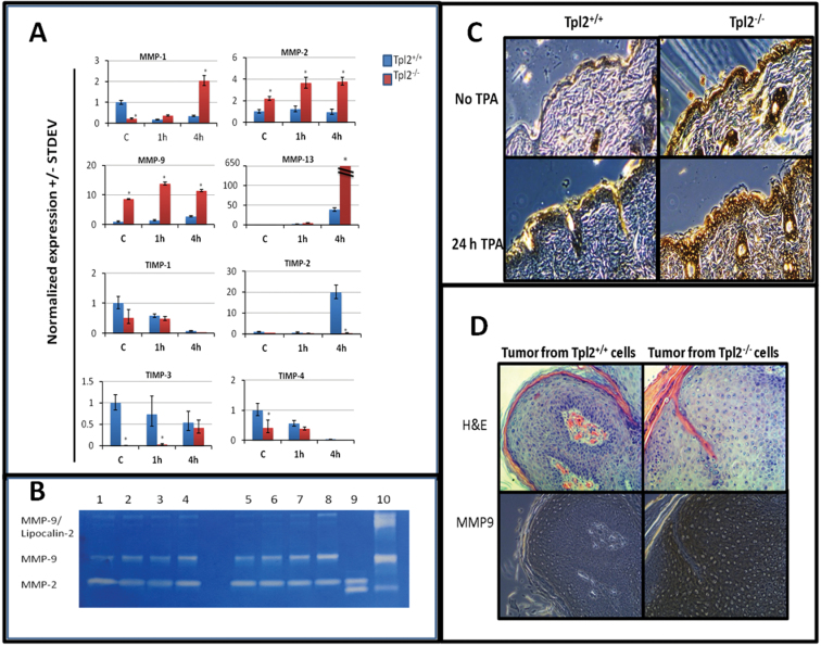
Fig. 2.
Tpl2 −/− primary keratinocytes have elevated gene expression of MMPs, reduced expression of Timp inhibitors and increased levels of active Mmp9 in vivo and in vitro. (A) qPCR results for Mmp1, 2, 9 and 13 and Timp1, 2, 3 and 4. Total RNA from basal- or TPA-stimulated Tpl2 +/+ and Tpl2 −/− keratinocytes was used for qPCR analysis. Note the broken graph for Mmp13 due to the extremely high gene expression in Tpl2 −/− keratinocytes. Asterisks denote significance at the P ≤ 0.001 level. (B) Gelatin zymography for Mmp2/Mmp9. Protein from conditioned media was electrophoresed and analyzed for expression of active Mmp2 and Mmp9. Lane 1: Tpl2 +/+ control; Lane 2: Tpl2 +/+ v-rasHa; Lane 3: Tpl2 +/+ TPA; Lane 4: Tpl2 +/+ v-rasHa + TPA; Lane 5: Tpl2 −/− control; Lane 6: Tpl2 −/− v-rasHa; Lane 7: Tpl2 −/− TPA; Lane 8: Tpl2 −/− v-rasHa + TPA; Lane 9: Mmp2-positive control; Lane 10: Mmp9-positive control. (C) Immunohistochemistry of Mmp9 in TPA-treated skin. Shaved WT and Tpl2 −/− mice were treated with TPA (10 μg) for 0 or 24h and analyzed for expression of Mmp9. (D) Immunohistochemistry of Mmp9 in grafted tumors. Tumors developed on nude mice with v-rasHa-transduced Tpl2 +/+ (left) or Tpl2 −/− (right) keratinocytes mixed with fibroblasts from the same genotype were analyzed for Mmp9 expression. Magnification = ×20.

