Fig. 4.
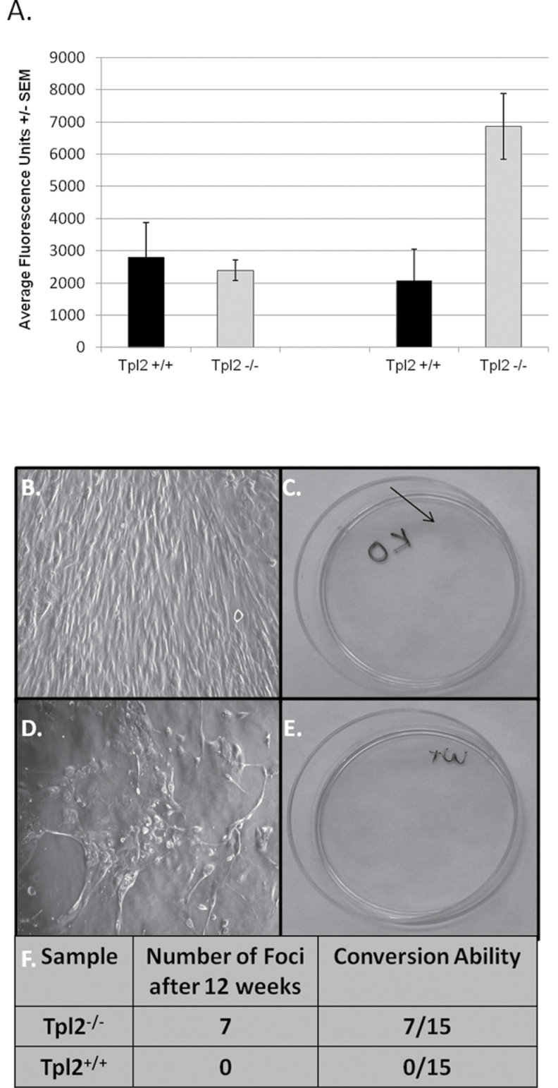
Tpl2 −/− primary keratinocytes infected with v-rasHa are more invasive than Tpl2 +/+ primary keratinocytes and undergo malignant conversion to a larger degree. (A) Invasion assay of v-rasHa-infected or vehicle-treated Tpl2 +/+ or Tpl2 −/− keratinocytes. Keratinocytes were allowed to invade Matrigel-coated inserts. Invasive cells passing through membrane were stained with calcein and fluorescence measured. (B–F) Conversion assay of v-rasHa-infected keratinocytes. The Tpl2 −/− dishes containing foci display a morphology resembling a transformed state (B) and stain pink with rhodamine (C). In contrast, the v-rasHa-infected keratinocytes in the Tpl2 +/+ dishes do not form foci (D) and therefore did not stain with rhodamine (E). The conversion rate in Tpl2 −/− and Tpl2 +/+ dishes (F).

